
What is Glaucoma?
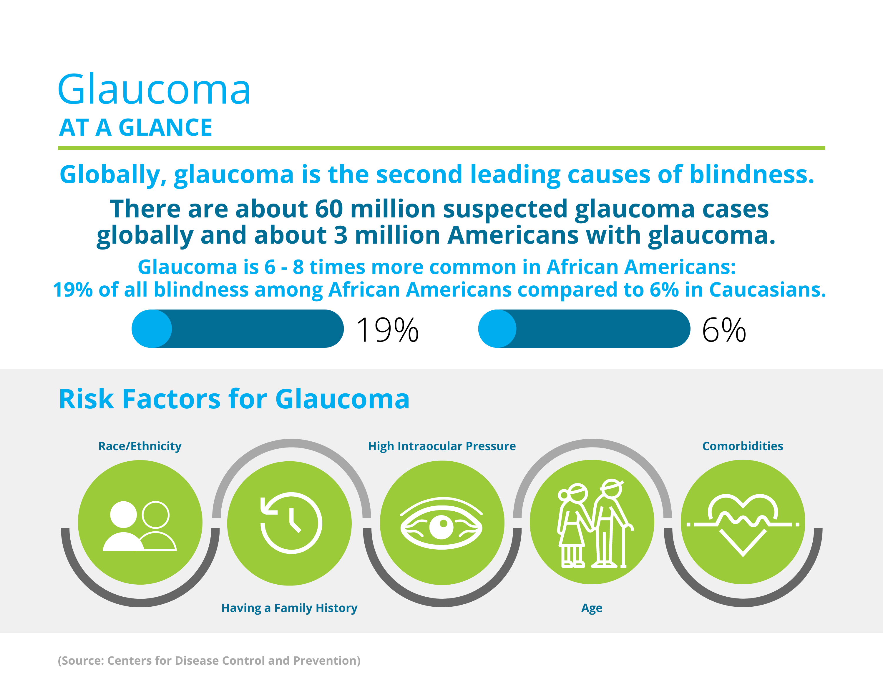
To understand glaucoma, it is important to take a step back and look at parts of the eye. In each eye there is an optic nerve. The optic nerve carries visual messages to the brain. Glaucoma occurs when there is damage to the optic nerve due to an increase in pressure, otherwise known as increased intraocular pressure. There are two main types of glaucoma:
Open-angle glaucoma: This is the most common form of glaucoma, this generally occurs after an increase pressure of a long period of time
Angle-closure glaucoma: Less common type of glaucoma, drainage may build up due to narrow angle in the eye
Causes?
Glaucoma occurs secondary to damage to the optic nerve. Elevated intraocular pressure (eye pressure) occurs due to a build-up of the fluid that flows and drains throughout the eye. Glaucoma can be hereditary, however that is not always the case. Some additional risk factors for glaucoma are older age, race, past medical history, eye injury or surgery, or corneas that are thin in the center.
Diagnosis & Treatment?
Generally the first diagnostic testing is a check on the pressure within the eye, usually done with a small pen that gently touches the surface of your eye after it is numbed with eye drops. In addition, a physician will most likely dilate eyes to have a better look at the optic nerve and structures within the eye. Dilation is done by a simple eye drop, generally causes no discomfort, and only lasts about 6 hours. A doctor may also test the thickness of your cornea using a method similar to checking the pressure. Imaging can be done to look at the optic nerve and evaluate for any swelling, as well as a visual field test to see if there is any decrease in vision.
Vision changes and eye damage due to glaucoma can’t be reversed, however there are treatment options to help slow down progression. Treatment options include medicated eye drops that a doctor will prescribe. If eye drops are not lowering pressure then oral medication or surgery may be needed.
Prevention?
Having regular dilated eye exams can help diagnose glaucoma in an early stage, and provide more effective treatment options. Talk with your eye doctor to find out how often you should be receiving dilated eye exams. If you are diagnosed with glaucoma, taking prescription eye drops and following doctor’s instructions will help prevent worsening of symptoms. In addition, if you are working with power tools, or doing activities where an eye injury may occur, wear protective eyewear.
For more information: https://www.nei.nih.gov/learn-about-eye-health/eye-conditions-and-diseases/glaucoma

What is Macular Degeneration?
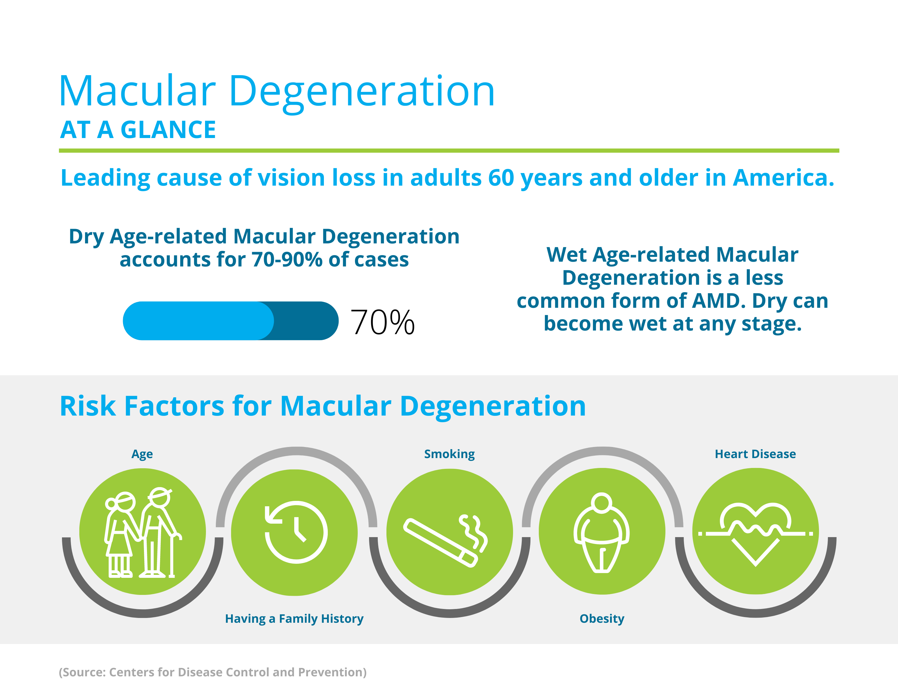
Age-related macular degeneration (AMD) happens due to damage to a part of the eye known as the macula. AMD is a common cause of severe vision loss in individuals over the age of 50. Early stages of macular degeneration may cause little to no symptoms. However, once AMD advances to more advanced stages then severe vision loss may occur. There are two types of macular degeneration:
Dry macular degeneration – the most common type. Vision loss with dry AMD is usually slow, and happens due to the slow breakdown of the macula.
Wet macular degeneration – less common type. Vision loss with wet AMD is often more severe, and happens due to abnormal blood vessels growing beneath the retina.
Causes?
There has been no specific cause of macular degeneration identified. However, there are some risk factors that may put an individual at a higher risk for developing AMD. One of the risk factors is family history, if your family has a history of AMD be sure to let your eye doctor know. Additional risk factors for developing AMD are smoking, high blood pressure, high cholesterol, obesity, being light-skinned, being female, light eye color, and eating a diet higher in saturated fat.
Diagnosis and Treatment?
Routine eye exams are used to diagnose macular degeneration. A doctor can see changes in your eyes during routine eye exams. During an eye examination, doctors may ask you to look at something called an Amsler grid. An amsler grid in a pattern of straight lines, if the lines are appearing wavy or missing then the doctor may do a more thorough exam. Doctors can also diagnostic imaging tests such as an OCT or fundus photos to evaluate changes in the eyes.
Treatment options for macular degeneration may help slow progression of AMD. However, this is no cure. Some doctors may choose to monitor condition if AMD is in its early stages. Usually this is in combination with a supplement of vitamins recommended by your doctor. There are some other treatment options that your doctor may recommend such as medication or laser therapy.
Prevention?
One way to help prevent the progression of AMD is by taking vitamin supplements. One of the most common combinations of supplements is vitamin C, vitamin E, zinc, lutein copper, and zeaxanthin. However, follow your doctor’s advice for the best supplement for you.
For more information: https://www.hopkinsmedicine.org/health/conditions-and-diseases/agerelated-macular-degeneration-amd


